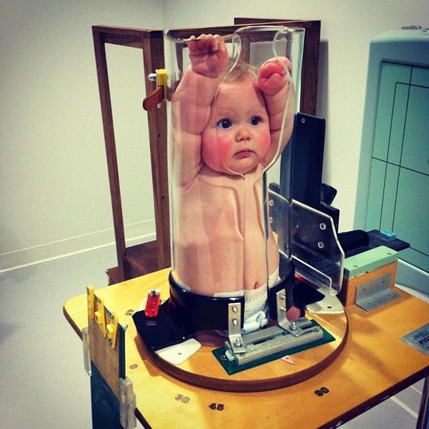
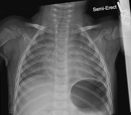
baby chest x ray technique
X-rays are forms of radiant energy like light or radio waves. This view is preferred in infant and neonate imaging whilst AP erect and PA erect views are ideal for children able to cooperate in sitting or standing 1.

Neonatal Chest Radiograph In The Exam Setting Radiology Reference Article Radiopaedia Org
The chest x-ray is performed to evaluate the lungs heart and chest wall.
. Chest X-rays can show a swallowed foreign object such as a coin. They can penetrate your body. The ROC-area was 080 in the derivation and validation sets.
They give your healthcare provider information about structures inside the body. Prevent hypothermia. While doing the X-ray Asepsis precautions.
Paediatric abdominal x-ray. As radiation protection is necessary for pediatric patients it is essential to image the chest properly and avoid unnecessary repeats. X-rays have more energy than rays of visible light or radio waves.
Chest pain or injury. It can detect signs of pneumonia a collapsed lung heart problems such as an enlarged heart and broken ribs or lung damage after an injury. A chest X-ray can help doctors find the cause of a cough shortness of breath or chest pain.
Children that present for abdominal x-rays are often very unwell therefore specialized techniques and appropriate communication are essential for gaining the childs cooperation. These tests expose children to low doses of radiation. In 202 87 patients from the derivation set a chest x-ray was performed.
The abdomen radiograph is a commonly requested examination in the pediatric patient. A normal chest x-ray could be predicted by increasing age increasing birth weight presence of rhinitis absence of retractions and increasing arterial oxygen saturation. In this study the exposure technique of 65 kVp and 16 mAs was chosen as a reference image due to this technique being near the suitable exposure uses in pediatric chest supine AP in the DR and.
A chest x-ray is typically the first imaging test used to help diagnose symptoms such as. X-rays are a kind of imaging test. A bad or persistent cough.
They can also help confirm that medical. Quieten the baby to avoid swings in respiratory depth While reading the X-ray Read schematically Do not jump to the diagnosis - you will miss important additional findings Make differential diagnosis and correlate clinically Write age in hoursdays on the X-ray. Pediatric Chest Screen 70-80 DIGITAL OPTIMUM kVp Universal CR Technique Chart using a standard 21 LgM Part View kV mAs kV mAs kV mAs Abdomen AP Grid 85 10 -15 85 20 - 25 85 30 - 40 Ankle AP 70 18 70 2 70 25 Ankle Obl 70 16 70 18 70 22 Ankle Lat 70 15 70 16 70 2 Chest -Adult AP 400 - tt -72 85 2 - 25 85 32 - 4 90 5 - 64.
If the pediatric patient can only manage a supine view this is more. Physicians use the examination to help diagnose or monitor treatment for conditions such as.

Ce4rt Guide For X Ray Techs To Immobilize Pediatrict Patients

Anterior Posterior A And Lateral B Chest X Ray Views Of The Cirs Download Scientific Diagram

Pediatric Chest Supine View Radiology Reference Article Radiopaedia Org

Chest Radiograph In Hospitalized Children With Covid 19 A Review Of Findings And Indications Sciencedirect

Pediatric Chest Horizontal Beam Lateral View Radiology Reference Article Radiopaedia Org

Diagnosis Of Other Lung Conditions In Premature Babies

Antarctica Neo Archive Que X Ray C Spine Ap View Image File No 0305 X Ray View Image History Of Science

Underinspiration And Poor Positioning Mimicking Lung Pathology In A Pediatric Patient Radiology Case Radiopaedia Org

An Ap Erect Chest Xray Of The Patient Showing An Upward Tilted Cardiac Download Scientific Diagram

The Normal Cxr Nurse Radiology Nursing Education

Chest X Ray Of The Neonate On First Day Of Admission Showing Multiple Download Scientific Diagram

Chest X Ray For Students How To Interpret And Present Methodically Andreas Astier


